strobeadmin

Home \ STROBE Admin

About STROBE Admin

This author has not yet filled in any details.
So far STROBE Admin has created 245 blog entries.

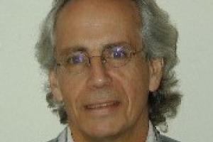
Margaret Murnane Presented Presidential Distinguished Service Award for the Irish Abroad

JILA Fellow Margaret Murnane was one of 10 recipients of the Presidential Distinguished Service Award for the Irish Abroad.

Tánaiste and Minister for Foreign Affairs Simon Coveney announced the names of the award winners on the 28th of November 2018. These awards, established in 2012, are meant to recognize the contributions of members of the Irish diaspora.

Each of the awards is for contributions to a specific category. Murnane received the science, technology and innovation award for her work as “one of the leading optical physicists of her generation”, according The Irish Times.

The awards were presented on Thursday, November 29th, 2018, by Ireland’s President Michael D. Higgens.

Other recipients of the award include Novelist Edna O’Brien and Irish-American Pulitzer Prize-winning author William Kennedy.

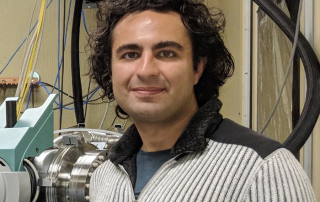
Congrats to Kasra Nowrouzi for Receiving the Spot Award from Lawrence Berkeley National Lab

Spot Award Purpose: The purpose of the Spot Recognition Award program is to acknowledge and reward outstanding individual and/or team workplace contributions that occur on a day-to-day basis. (For safety-related recognition, please see the Safety Spot Award Program Guidelines). Contributions should impact the quality, cost, service, safety, or resource utilization of an organizational unit, team, or department.

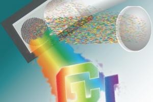
STROBE Tutorial: Point Spread Function Engineering

The point spread function describes fundamentally the function of an optical system. In this tutorial, we will present approaches for tailoring the response of an optical system for processing information at the sensor level and to enhance the overall performance of task specific imaging systems. In particular, we will introduce methods to shape the point spread function in three dimensions and their application in computational imaging.

Congrats to Margaret Murnane for Receiving the Presidential Distinguished Service Award

The Tánaiste and Minister for Foreign Affairs and Trade, Mr Simon Coveney, T.D., today announced the names of the recipients of the Presidential Distinguished Service Award for the Irish Abroad for 2018.

The Presidential Distinguished Service Award was established by the Government following the 2011 Global Irish Economic Forum as a means to recognise the contribution of members of the Irish diaspora, and the first awards were made in 2012.

Announcing the recipients of the award, the Tánaiste said:

“I am delighted to have the opportunity once again this year to formally recognise the achievements of some of the finest members of our global family, our diaspora.”

“The diverse contributions of these individuals extends across six different categories. Through their work, they contribute to building an invaluably positive image of Ireland and Irish people around the world.”

“This is the seventh year in which recipients have received this Award, and the ceremony remains a very important opportunity to acknowledge those who have contributed to realising Ireland’s place as an island at the centre of the world.”

Novelists Edna O’Brien and William Kennedy among winners of award for Irish abroad

Limerick-born Prof Margaret Murnane will be given the award for the science, technology and innovation award, which will be presented to her in the US. Prof Murnane is regarded as being one of the leading optical physicists of her generation. She is Director of the National Science Foundation STROBE Science and Technology Center on functional nano-imaging, a fellow at JILA and Distinguished Professor at the Department of Physics and Electrical and Computer Engineering at the University of Colorado.

Paper on diffractive optics shows 3D holograms become reconfigurable by computer

Diffraction refers to a variety of phenomena occurring when a wave encounters an obstacle and bends around it. Diffractive optics are widely used today in imaging, holography, microscopy and manufacturing. Previous work has shown that extending diffractive optics from two dimensions to three dimensions enables new functionality and improves system performance. The paper suggests a way to make the two-dimensional waves three-dimensional in real time with a simple modification to existing devices controlled with a computer.

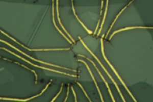
Super-Resolution Imaging of Clickable Graphene Nanoribbons Decorated with Fluorescent Dyes

A STROBE team led by Ke Xu at Berkeley adapted advanced super-resolution microscopy techniques developed by the Weiss group at UCLA, to rapidly image graphene nanoribbons (GNRs) and carbon nanotubes (CNTs). These are important functional materials with great promise in nano-electronics and other applications. However, it has been a challenge to visualize and characterize them in a high-throughput manner due to their small physical dimensions. Temporal fluorescence intensity fluctuations in these samples enabled the reconstruction of SRM images through superresolution optical fluctuation imaging (SOFI) and the related superresolution radial fluctuation (SRRF) analysis methods.

Sub-ångström Cryo-EM Structure of a Prion Protofibril Reveals a Polar Clasp

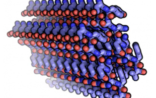
Brain dysfunction can be caused by aggregated proteins such as prions. In humans, the prion protein causes an infection by changing its shape and transforming into rope like aggregates. In this way, stable prion aggregates are immune to normal processes of destruction in an organism and bring about neuronal death and ultimately disease. There is no molecular explanation for the variable efficiency of prion spread between species. To investigate this phenomenon, STROBE scientists Rodriguez and Maio used cryo electron microscopy to reveal the atomic structures of regions within prion that have a high propensity to form structured aggregates. This made it possible to take an atomic look at structural motifs that may represent the molecular basis for a prion species barrier.

Demonstration of Electron Ghost Imaging

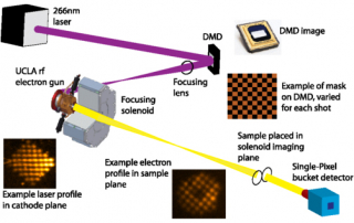
The first demonstration of computational ghost imaging with electrons has been carried out at UCLA. A digital micromirror device is used to directly modulate the photocathode drive laser to control the transverse distribution of a relativistic electron beam incident on a sample. Correlating the structured illumination pattern to the total sample transmission then retrieves the target image, avoiding need for a pixelated detector. In our example, we use a compressed sensing framework to improve the reconstruction quality and reduce the number of shots compared to raster scanning a small beam across the target. Compressed electron ghost imaging can reduce both acquisition time and sample damage in experiments for which spatially resolved detectors are unavailable (e.g. spectroscopy) or in which the experimental architecture precludes full frame direct imaging.

Go to Top