strobeadmin

Home \ STROBE Admin

About STROBE Admin

This author has not yet filled in any details.
So far STROBE Admin has created 245 blog entries.

Study reveals key details about bacterium that increases risk for stomach cancer

More than half of the people in the world host colonies of a bacterium called Helicobacter pylori in their stomachs.

Although it’s harmless to many, H. pylori can cause stomach cancer as well as ulcers and other gastric conditions. Doctors tend to prescribe multiple antibiotics to defeat the microbe, but that strategy can lead to antibiotic-resistant superbugs.

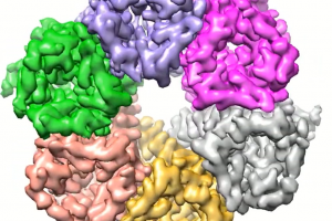
Now, a finding by UCLA scientists may lead to a better approach. The researchers have determined the molecular structure of a protein that enables H. pylori to stay alive in the stomach, and elucidated the mechanism by which that protein works.

Z. Hong Zhou, the study’s corresponding author and a UCLA professor of microbiology, immunology and molecular genetics, said the findings answer questions that have been sought ever since 2005, when two Australian scientists won a Nobel Prize for their discovery of H. pylori and its role in gastritis and peptic ulcer disease.

Innovation in the Semiconductor Industry: Implications for Education and Diversity

Dr. Laura Oliphant has been at the forefront of technology for over 25 years, first at Intel, as an Engineer and Corporate Venture Capitalist, and now, as a General Partner of a Venture Fund, Board Director and CEO of startups.  She will talk about her career trajectory, the challenges for technology innovation going forward, the implications for education, diversity, and what she wishes that she had known in starting out in her career.

Laura is a General Partner with Spirit Ventures, a new venture capital firm that will focus on key enabling technologies. She is an experienced CEO, Board Member, and Investor who was an Investment Director in Intel Capital from 2001 until her retirement from Intel in 2016. At Intel, she brought significant strategic and financial value to Intel, and she was awarded Intel’s highest award, the Intel Achievement Award, for her contributions. After retiring from Intel, she was the CEO of Translarity, an investor backed, semiconductor test startup. Laura is also currently part of the Board of Directors for Thin Film Electronics ASA (THIN), a public company, Novelda AS, and Numascale AS, all based in Oslo, Norway. Prior to her role in Intel Capital, Laura served as a Supply Chain Program Manager in Intel’s Technology and Manufacturing Group (TMG). Laura was one of the key coordinators for Intel’s transition to the 300 mm wafer size in their factories, a project which delivered to cost targets and added to Intel’s gross margin. Laura was also the co-chairperson of the SEMATECH Metrology and Yield Management Advisory Group, and was part of the International Technology Roadmap for Semiconductors (ITRS) committee for yield management technology. Laura is currently on the board of advisors for the UC Berkeley Skydeck Accelerator and has served on the Lawrence Berkeley Lab Innovation Grant Committee. Laura received her PhD in Chemical Engineering from the University of California, Berkeley, where her thesis research was centered on candidate batteries for electric cars.

3D Phase Contrast Tomography with Atomic Resolution

Electron tomography is a technique used in both materials science and structural biology to image features well below optical resolution limit. In this work, we present a new algorithm for reconstructing the three-dimensional(3D) electrostatic potential of a sample at atomic resolution from phase contrast imaging using high-resolution transmission electron microscopy. Our method accounts for dynamical and strong phase scattering, providing more accurate results with much lower electron doses than those current atomic electron tomography experiments. Our simulation results show that, for a wide range of experimental parameters, we can accurately determine both atomic positions and species, and also identify vacancies even for light elements such as silicon and disordered materials such as amorphous silicon dioxide and also identify vacancies. Our preliminary experimental results also show promising outcome of the method.

Best Practices in Science Communication

In this session of the STROBE seminar, we go through best practices of visual communication and then workshop materials brought by the participants (you!). Participants sent a maximum 1-slide document to Ellen and Nico before the seminar. We break out into groups at each node to workshop the materials as a group, with input from experienced communicators. The material can be intended for your 3-minute thesis presentation, or for a conference presentation.

Leveraging the Extreme Ultraviolet: Extending Correlative Microscopy into Industrial Applications, the Time Domain, and More

In the last century and defining the first two decades of this one, the development of novel materials and manufacturing processes has demanded the advancement of new characterization techniques. This characterization leveraged light in its many rich forms: While optical probes proved tractable in the first half of this timeframe, it took the emergence of synchrotrons and other X-ray sources and optics to penetrate matter and move to higher photon energies. Only in the very recent past, however, have innovators been able to successfully utilize the Vacuum region of the spectrum (VUV, EUV, and Soft X-ray) effectively in the laboratory. This long-overlooked region of the spectrum is proving to be a rich and promising probe for practical materials and devices—filling a void in the existing characterization and imaging space.

In this talk, we will discuss the advent of Coherent EUV light as the next technique to complement this correlative suite of instruments. Uniquely merging diffractive imaging, spectroscopic, and time-resolved measurements enables key applications that will unlock new avenues ranging from Semiconductor metrology, fundamental materials and device characterization. We will discuss how it takes a diverse and extensive team to bring such technology from an idea to impact. More importantly, we will discuss the evolution of a technology that has been taken from the limited confines of the Synchrotron community to very soon becoming a laboratory instrument available to augment the rich tool suite now relied upon by academic researchers and industrial microscopists alike.

Elemental distribution and local electronic structure in oxynitride nanocrystals

Zinc-gallium oxynitride (Ga1-xZnx)(N1-xOx) exhibits visible absorption with a band gap that depends on composition (i.e., the value of x) and has been demonstrated to split water under visible irradiation. The origin of visible absorption in this solid solution material in this material is not understood. Furthermore, the local atomic-level distribution of the 4 elements, Ga, Zn, N, and O may play an important role in the optical properties of (Ga1-xZnx)(N1-xOx).

This presentation will focus on our use of scanning transmission electron microscopy (STEM) tools to characterize the local composition and electronic structure of these particles. I will describe the elemental distribution within (Ga1-xZnx)(N1-xOx) nanocrystals, measured by Energy Dispersive X-ray Spectroscopy (EDS) with sub-nm resolution, as well as the methods to control compositional disorder. Furthermore, I will describe our ongoing efforts to use Electron Energy Loss Spectroscopy (EELS) to correlate local composition with local electronic structure and elucidate the relationship between the two. Together, these tools allow us to postulate a comprehensive picture of the optical properties of (Ga1-xZnx)(N1-xOx) nanocrystals.

Atomic cryoEM for proteins, nucleic acids and lipids

Cryo electron microscopy (cryoEM) has emerged as a tool of choice for determining three-dimensional (3D) structures of macromolecular complexes or biological nano-machines (>50 kDa) in their native forms. When such complexes can be isolated in microgram quantities, atomic models can now be obtained by cryoEM single-particle analysis and model building. Comparisons of atomic models obtained for the same complex at different functional states provide mechanistic insights for its functions. For pleomorphic complexes, such as those in their cellular or tissue environments, molecular resolution structures can be reconstructed by cryo electron tomography (cryoET). Examples will be presented to illustrate the power of cryoEM in visualizing 3D structures of nano-scale biological machines containing proteins, nucleic acids or lipids to inform such fundamental biological processes as genome transcription, molecular translocation and infectious diseases.

Congrats to Jessica Ramella-Roman for Being Elected as a 2019 SPIE Fellow

Each year, SPIE promotes Members as new Fellows of the Society. SPIE will honor 88 new Fellows of the Society this year. Fellows are Members of distinction who have made significant scientific and technical contributions in the multidisciplinary fields of optics, photonics, and imaging. They are honored for their technical achievement and for their service to the general optics community and to SPIE in particular. More than 1,400 SPIE members have become Fellows since the Society’s inception in 1955. Dr. Jessica Ramella-Roman, Florida International University, United States was elected for achievements in spectro-polarimetric techniques for diagnostic applications.

Congrats to Jose Rodriguez for Being Selected as One of C&EN’s Talented Twelve

Welcome to the fourth annual Talented 12 issue. We’ve spent months searching high and low for these bright, young scientific minds and are excited to finally introduce you to them. Here, you’ll meet a dozen chemists pushing the boundaries in their fields. To say they are tackling life’s great mysteries would be an understatement. The interests of these distinguished researchers include deciphering the chemistry that enabled life on Earth, exploring molecules in far-flung parts of our solar system, designing out-of-this-world materials that can store energy or mimic human organs, and developing technology to precisely alter the code of life.

Go to Top