strobeadmin

Home \ STROBE Admin

About STROBE Admin

This author has not yet filled in any details.
So far STROBE Admin has created 245 blog entries.

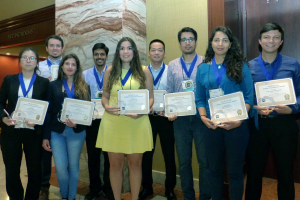
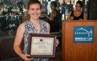
Congrats to Jessie Woodcock for Receiving an Outstanding 2018 STEM Partner Award in recognition of partnership and support of Workforce Development & Education programs

On Thursday, September 26, Workforce Development & Education hosted our annual Mentor Appreciation event where we recognized our outstanding mentors and STEM partners. This event highlighted accomplishments for FY2018. Outstanding 2018 STEM Partner is hereby awarded on this 26th day of September 2019, to Jessie Woodcock, in recognition of partnership and support of Workforce Development & Education programs.

Congratulations to Josh Knobloch for receiving a 2019 TECHCON Student Presentation Award

Thank you to the SRC students, industry, and faculty that attended TECHCON and made it a great success. The final event for TECHCON 2019 was presenting Top 10 Student Presentation Awards and the URI Best Poster Awards at Tuesday’s Dinner.

2019 TECHCON Student Presentation Award Winner:

Joshua Knobloch
Nanoscale Metrology and Imaging of Layered and Nano-enhanced Materials using Coherent Extreme Ultraviolet Beams

Webinar: FRIPS Science Communication

Front Range Industry Postdoc Summit Science Communication Webinar, Fall 2019. Learn important tips and tools to distill your research for any audience. In this webinar, we teach you what science communication is, why it’s important for your career, and tools to help you create an adaptable elevator pitch understandable to any audience.

STROBE Responsible Conduct of Research Seminar

You must be a STROBE member to access this section of the website. To get access to Seminar Videos, Publication PDFs, Career Opportunities and more, request a STROBE membership.


If you are already a STROBE member, log in and then return to this page.

Watching crystal nucleation happen at atomic scale

Crystals form in storm clouds, metals, drug molecules, and even in diseased tissues. Despite their ubiquity, scientists still don’t fully understand what happens when a liquid solution first starts to form a solid crystal, a step called nucleation. Now researchers have gotten their first glimpse of the details of the process, imaging individual atoms during nucleation in metal nanoparticles (Nature 2019, DOI: 10.1038/s41586-019-1317-x).

First 4D look at crystallising atoms contradicts textbook nucleation theory

For the first time scientists have watched iron and platinum atoms crystallise in 4D – not only observing their arrangement in space but tracking them over time. Their observations clash with classical nucleation theory, which describes the early stages of a phase transition, adding to growing evidence that the textbook theory is outdated and imprecise.

Atomic motion is captured in 4D for the first time

Results of UCLA-led study contradict a long-held classical theory.

Everyday transitions from one state of matter to another — such as freezing, melting or evaporation — start with a process called “nucleation,” in which tiny clusters of atoms or molecules (called “nuclei”) begin to coalesce. Nucleation plays a critical role in circumstances as diverse as the formation of clouds and the onset of neurodegenerative disease.

A UCLA-led team has gained a never-before-seen view of nucleation — capturing how the atoms rearrange at 4D atomic resolution (that is, in three dimensions of space and across time). The findings, published in the journal Nature, differ from predictions based on the classical theory of nucleation that has long appeared in textbooks.

Real Time Near-field Imaging of Biological and Nano-systems

Label-free chemical nano-imaging in dense molecular environments has remained a long-standing challenge. STROBE Thrust lead Markus Raschke led a team of academic and national laboratory scientists from LBNL, Boulder and Berkeley to speed-up scanning near-field optical microscopy (s-SNOM) by a factor of 10! This remarkable achievement allowed the team to image the surface shape and chemistry of biological samples, including mollusk shells, with nanometer spatial resolution.

De-blurring Images of Living Biological Samples

Imaging fast moving samples such as living biological samples is challenging because the images can appear blurred if the strobe light is not fast enough. This is an issue for high-quality quantitative phase imaging, which was too slow for many samples (image on right). STROBE faculty Laura Waller from Berkeley led a team to develop a new approach to reduce this blur, that enhances the speed to match the frame rate of the detector, to enable much clearer imaging of many biological systems (image on left).

Electron Imaging Reveals the Conductance Watershed Between Two Electrodes

Traditional transmission electron microscopy (TEM) excels at determining the physical structure of a sample, but reveals little about the electronic structure. STROBE faculty Chris Regan at UCLA developed a new technique based on secondary electron emission (SE) and electron beam induced current (EBIC) to image the electronic structure of functioning devices with a TEM-like spatial resolution. He used this new SEEBIC technique to map the conductance of a nanodevice containing a thin silicon membrane (brown and green) separating two electrodes (blue).

Go to Top