Research Highlights

Home \ Research \ Research Highlights

Imaging Material Functionality Through Three-dimensional Nanoscale Tracking of Energy Flow

April 28, 2020|

The next generation of semiconducting materials that will facilitate energy transport and storage in the technologies around us is becoming increasingly complex. The ability of energy carriers to move between atoms and molecules underlies biochemical and material function. Understanding and controlling energy flow, however, requires observing it on ultrasmall and ultrafast spatio-temporal scales, where energetic and structural roadblocks dictate the fate of energy carriers.

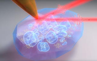
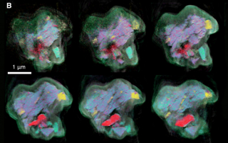
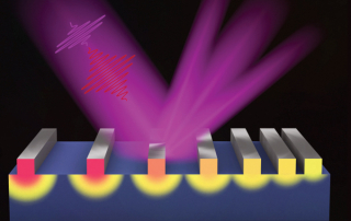
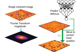
A STROBE team led by Naomi Ginsberg (UCB) developed a novel time-resolved interferometric scattering microscope to visualize how energy navigates the intrinsically disordered landscapes in these materials on the nanoscale. With this high-throughput technique, they collected non-invasive stroboscopic movies in a variety of organic, inorganic, and hybrid materials to demonstrate its powerful versatility. Applied to other cutting-edge materials, we hope to inform the design of new functional devices for the semiconductor industry of tomorrow.

Delor, M. et al., "Imaging Material Functionality Through Three-dimensional Nanoscale Tracking of Energy Flow," Nature Materials, 19, 56 (2020).

Understanding the Role of Molecular Disorder in Organic Electronics and Photonics

April 26, 2020|

How molecules interact and transfer energy between each other dictates the performance in molecular electronics, organic light emitting diodes, photovoltaics, or in many biological processes. However, imaging the controlling underlying molecular order and associated wavefunction delocalization on the molecular scale has long remained a major challenge in imaging science.

A STROBE team from CU Boulder, UC Berkeley and LBNL, has overcome this challenge developing a new technique of nanoimaging in the infrared probing the delicate low-energy landscape of molecular interactions. Measuring coupled molecular vibrations with high precision provides for a new molecular ruler to resolve the effect of disorder with sub-nanometer resolution. In a representative organic electronic material of metal-porphyrin nano-crystals the researchers learned about the relationship between structure and function of energy transfer on molecular length scales. The new insights gained advance our understanding of light harvesting in photosynthesis and improve the design of next generation organic electronic and photonic devices.

Muller et al., “Vibrational exciton nanoimaging of phases and domains in porphyrin nanocrystals” PNAS, 117, 7030 (2020).

Multimodal X-ray and Electron Microscopy of the Allende Meteorite

September 20, 2019|

A STROBE team from UCLA, Berkeley and Boulder developed a nanoscale multimodal X-ray and electron microscopy framework that is applicable to a wide range of inhomogeneous samples with complex structural and chemical properties. Using an Allende meteorite as an example, we performed structural and chemical mapping to infer the mineral composition and its potential processes. This work opens a route to future microscopies of complex materials.

Y.Hung Lo et al., “Multimodal x-ray and electron microscopy of the Allende meteorite”, Science Advances 5, eaax3009 (2019).

Atomic Motion Captured, for the First Time, in 4D

June 26, 2019|

Everyday transformations from one state of matter to another—such as freezing, melting or evaporation – start with a process called “nucleation”, in which tiny particles containing just a few atoms or molecules begin to coalesce. Nucleation plays a critical role in events as diverse as the formation of clouds and the onset of neurodegenerative disease. STROBE Deputy Director Jianwei (John) Miao, led an interdisciplinary team from Lawrence Berkeley Lab, University of Colorado Boulder, University of Buffalo and the University of Nevada Reno, to gain a never-before-seen view of nucleation—capturing how the atoms rearrange in the tiny seed particles at atomic resolution. Their findings, published in the journal Nature, differ from predictions based on the classical theory of nucleation that has long appeared in textbooks.

J. Zhou et al., "Observing crystal nucleation in four dimensions using atomic electron tomography," Nature 570, 500-503 (2019).

Stroboscopic Imaging of Nanoscale Transport

June 14, 2019|

The functional properties of photovoltaics and nano-devices for electronics, thermoelectrics and data storage can be enhanced by tuning their structure at the nanoscale. However, at dimensions <100nm, bulk models can no longer accurately predict heat, charge or spin transport, or the mechanical properties of doped or nano-structured materials. A STROBE team from CU Boulder, UC Berkeley, and LBNL developed a real-time microscope to capture, map and understand nanoscale heat transport in nanostructures. This microscope was then used to validate a very surprising prediction—that an array of closely-spaced nanoscale heat sources can cool more quickly than when spaced far apart.

Frazer et al., “Engineering nanoscale thermal transport: Size- and spacing-dependent cooling of nanostructures,” Physical Review Applied 11, 024042 (2019); Karl et al., “Full-Field Functional Imaging of Nanoscale Dynamics Using Tabletop High Harmonics”, Science Advances 4, eaau4295 (2018).

Deep Learning for Single-Shot Autofocus Microscopy

June 5, 2019|

A STROBE team led by Prof. Waller, created a novel computational imaging autofocusing system for microscopes utilizing an of-the-shelf LED and a machine learning algorithm with optical physics knowledge incorporated into its design.

H. Pinkard et al., “Deep learning for single-shot autofocus microscopy”, Optica 6, 794 (2019).

Information-rich Localization Microscopy Through Machine Learning

April 30, 2019|

Artificial neural networks enable the extraction of multiple parameters, including spectral and depth information, from unmodified experimental single-molecule images for multidimensional super-resolution microscopy. Good color separation is thus achieved in fixed cells using two dyes ~80 nm apart in emission wavelength.

T. Kim et al., “Information-rich localization microscopy through machine learning”, Nature Communications 10, (2019).

Real Time Near-field Imaging of Biological and Nano-systems

April 3, 2019|

Label-free chemical nano-imaging in dense molecular environments has remained a long-standing challenge. STROBE Thrust lead Markus Raschke led a team of academic and national laboratory scientists from LBNL, Boulder and Berkeley to speed-up scanning near-field optical microscopy (s-SNOM) by a factor of 10! This remarkable achievement allowed the team to image the surface shape and chemistry of biological samples, including mollusk shells, with nanometer spatial resolution.

MS. C. Johnson et al., "Infrared nanospectroscopic imaging in the rotating frame," Optica 4, 424 (2019).

Temporal magnification for streaked ultrafast electron diffraction and microscopy

January 29, 2019|

UCLA STROBE researchers show how a radiofrequency cavity can be used as an electron longitudinal lens in order to produce a highly magnified temporal replica of an ultrafast process, and, in combination with a deflecting cavity, enable streaked electron images of optical-frequency phenomena, taking advantage of the time-stretch concept.

D. Cesar et al., “Temporal magnification for streaked ultrafast electron diffraction and microscopy” , Ultramicroscopy 199, 1 - 6 (2019).

De-blurring Images of Living Biological Samples

December 10, 2018|

Imaging fast moving samples such as living biological samples is challenging because the images can appear blurred if the strobe light is not fast enough. This is an issue for high-quality quantitative phase imaging, which was too slow for many samples (image on right). STROBE faculty Laura Waller from Berkeley led a team to develop a new approach to reduce this blur, that enhances the speed to match the frame rate of the detector, to enable much clearer imaging of many biological systems (image on left).

M. Chen et al., "Quantitative differential phase contrast (DPC) microscopy with computational aberration correction," Optics Express 26, 32888-32899 (2018).
Go to Top