Atomic structures determined from digitally defined nanocrystalline regions
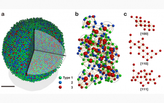
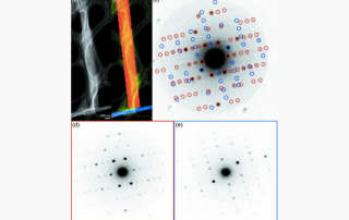
Three-dimensional (3D) structures of molecules determined from nanoscale regions of crystalline arrays could potentially illuminate the subtle differences that engender crystal defects or the multiple states accessible to subpopulations of molecules within an ensemble. A step toward this goal involves the extraction of meaningful diffraction data from 3D regions on the nanoscale. This is achieved using a near-parallel electron beam designed to illuminate sub-10nm regions of a sample. Scanning such a beam across a sample allows for digital logic to be applied to the measured data, facilitating the expostfacto assortment of information and reduction from desired 3D subvolumes.
A STROBE team from UCLA, UC Berkeley and LBNL collaborated to determine the first molecular structures determined by 4DSTEM. The structures were determined from a digitally defined subregion of a nanocrystal. After collecting TB of data, the team obtained reconstructions that revealed the atomic structure of a peptide, and showed that radiation damage imparted on the sample during data collection was not prohibitive for structure determination. Compared to other approaches, the approach allows for a much greater degree of control and obviates the need for spatial separation of samples. New methods, algorithms, enhanced microscopes and advanced sample preparation techniques developed by the STROBE collaboration were key to enabling the success of this project.