Congrats to Ke Xu for Being Selected as a Pew Innovation Fund Investigator
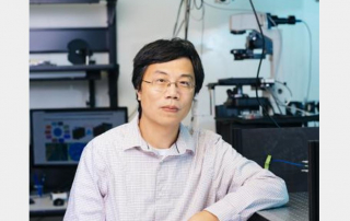
The Pew Scholars Program in the Biomedical Sciences provides funding to young investigators of outstanding promise in science relevant to the advancement of human health. The program makes grants to selected academic institutions to support the independent research of outstanding individuals who are in their first few years of their appointment at the assistant professor level. Congratulations to Ke Xu for being selected as a Pew Innovation Fund Investigator!