masonlw

Home \ Lauren Mason

About Lauren Mason

This author has not yet filled in any details.
So far Lauren Mason has created 340 blog entries.

High-Resolution Broadband Ptychography with a Tabletop EUV Continuum

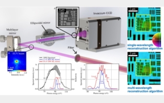
Ptychography implemented with coherent high-harmonic (HHG) sources enables high-resolution, high-fidelity imaging of nanostructures and biosystems. However, when driven by mid-infrared lasers to generate light at higher photon energies, HHG inherently produces a broadband quasi-continuum, which is less suited for coherent imaging compared with a single harmonic order. Consequently, experiments typically select a narrow bandwidth of ≈1%, leaving most of the HHG photons unused, increasing exposure times. In this work, we demonstrate broadband ptychography utilizing an extreme UV (EUV) continuum centered at 92 eV, with a bandwidth of up to 7.9 eV (a relative bandwidth of ~9%). By focusing the HHG beam to a sub-micrometer spot size to relax the temporal coherence constraints, and utilizing a multi-wavelength ptychographic reconstruction algorithm, we achieve a spatial resolution of 42 nm, which is near the diffraction limit of ~30 nm for our setup. To the best of our knowledge, this represents the broadest spectral bandwidth successfully employed to date for EUV ptychography, with the potential to increase the usable photon flux by up to an order of magnitude relative to previous approaches. In the future, broadband soft X-ray ptychography can be used to image hydrated samples around the carbon K-edge and magnetic textures at the L-edges of transition metals.

Imaging the functioning of FeFETs

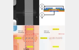
Ferroelectric materials such as hafnium zirconium oxide (HZO) are likely to form the basis of a next-generation memory technology.  For instance, replacing the conventional gate dielectric in a field-effect transistor (FET) with HZO can form a ferroelectric FET, or FeFET.  Chris Regan’s group at UCLA has been working with Suman Datta’s group at Georgia Tech to speed development of this exciting technology.  Standard scanning transmission electron microscopy (STEM) (see top left figure), shows the basic layout of a device fabricated at Georgia Tech, but it reveals little about its function.  STEM electron-beam induced current (EBIC) imaging at UCLA, on the other hand, shows exactly how the FeFET works.  With two EBIC transimpedance amplifiers (Fig. X, top right), the STROBE team can form two linear combinations.  The STEM EBIC sum image (see lower left figure), gives secondary-electron EBIC (SEEBIC) contrast. It maps the conductors, correctly showing that the electrode in the upper left corner is not connected to the gate, source, or drain. The STEM EBIC difference image (see lower right figure) gives electron-hole-pair separation contrast, and it is even more informative. It reveals the electric fields that control the FeFET’s function: the depolarization fields in the ferroelectric HZO where the gate electrode overlaps the semiconducting indium tungsten oxide (IWO).  Moreover, it reveals these fields just as well whether it has to image through 40 nm of palladium or not. Higher magnification images of the FeFET in both polarization states (not shown) show exactly how much of the ferroelectric switches, providing a structural explanation of the FeFET threshold voltages and memory window. This functional imaging technique – STEM EBIC imaging – is a unique and uniquely powerful development enabled by STROBE.

Trypanosome doublet microtubule structures reveal flagellum assembly and motility mechanisms

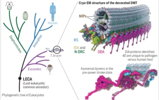
Integrated cryogenic electron microscopy (cryoEM) and cryogenic electron tomography (cryoET) approach, advanced through NSF’s STROBE, a National Science Foundation Science and Technology Center, enables unparalleled resolution for complex cellular structures like the flagellar doublet microtubules (DMT) of Trypanosoma brucei. This parasite causes African sleeping sickness, affecting millions. The flagellum is essential for motility, infectivity, and survival across different host environments. STROBE-supported cryoEM at the EICN resource at UCLA allowed high-resolution imaging of native structures, bypassing artifacts of recombinant systems and revealing detailed architecture of the 96-nm repeat with 154 proteins, including 40 parasite-specific ones (see figure).

The study provides critical insights into motility mechanisms by capturing axonemal dynein motors in the pre-power stroke conformation. This supports a novel ‘dragon boat’ model in which coordinated power strokes of dyneins drive microtubule sliding, generating the characteristic helical waveform that powers parasite movement through blood and tissues. Significantly, dyneins are the molecular motor across all living cells, including ours, and the mechanisms are universal.

This STROBE-supported, NSF-funded cryoEM study elucidates the mechanochemical basis of flagellar motility, offering new avenues for therapeutic intervention against trypanosomatid diseases and advancing our understanding of eukaryotic flagella evolution and function.

Designing lensless imaging systems to maximize information capture

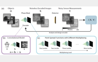
Lensless imagers can make cameras far thinner than conventional optics and can compress rich scene information into a single measurement. But the same optical multiplexing that enables those advantages can also scramble a scene so strongly that reconstruction loses the information it needs. We address this design problem by evaluating lensless encoders through mutual information estimated directly from noisy measurements. By combining a probabilistic model of measurement distributions with a detector-noise model, we compare encoders without tying performance to any one reconstruction algorithm and quantify how object sparsity, encoder multiplexing, and noise interact. Across simulations, we show that dense objects are best matched to low-multiplexing encoders, whereas progressively sparser objects benefit from higher multiplexing. We then optimize phase-mask encoders for specific object classes, and these information-optimal designs outperform heuristic masks in mutual information while also improving downstream reconstruction quality. Experiments with a conventional lens, a random multi-focal lenslet array, and a diffuser reinforce the same lesson for dense natural images: more multiplexing can reduce recoverable information. By making information capture itself the design target, our work offers decoder-independent engineering rules for lensless imaging and other multiplexed computational imaging systems. Because the best encoder depends on the assumed object distribution and mutual information does not guarantee every task-specific outcome, the next step is to test robustness under distribution shift, alternate noise models, and specialized downstream tasks.

Multimodal Nanoscale Mapping of Local Structure and CO2 Adsorption in Metal–Organic Frameworks

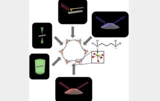
Currently, host-guest interactions in metal–organic frameworks (MOFs) are mainly understood via theoretical simulations, bulk characterization techniques, and x-ray diffraction, all of which can average out information related to defects, interfaces, and spatial heterogeneity among adsorption sites. Since these structural features can directly impact the functionality of MOFs we applied local probes to investigate these effects.  Specifically, we applied both 3D electron diffraction and 4D scanning transmission electron microscopy to structurally characterize and spatially visualize the effect of gas-loading and diamine functionalization in MOF crystals. In addition, complementary infrared scattering scanning near-field optical microscopy measurements confirmed spatial variations of chemisorbed CO2 inside an individual crystal.

The results of our multimodal approach was the ability to map heterogeneity arising from host-guest interactions and material functionalization.  Our 3D electron diffraction data has provided the only ab initio electrostatic potential map of any variant of Mg2(dobpdc), which has been an elusive structure. Our 4D-STEM images are, to our knowledge, the only dual-space (i.e., real and reciprocal space) visualizations of a MOF. Most importantly, although we have applied this methodology to a specific material of interest in gas storage and separation, we hope to introduce these microscopy techniques to the community as effective tools, complementary to the established bulk techniques, by which to evaluate and optimize synthetic and post-synthetic modification procedures of MOFs.

Accurate determination of the 3D atomic structure of amorphous materials

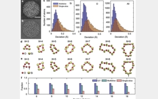
Amorphous materials — solids lacking long-range periodic order — are vital to technologies spanning thin-film electronics, solar cells, phase-change memory, magnetic components, biomedical devices, and quantum hardware. Yet their structural disorder has long prevented full three-dimensional (3D) atomic mapping. Despite major advances in probing short- and medium-range order, experimentally determining complete 3D atomic arrangements in amorphous solids remains challenging. Atomic electron tomography (AET) now offers a route to direct 3D atomic imaging without requiring crystallinity or averaging. In a forthcoming Nature paper, Miao and collaborators present a quantitative analysis of AET, showing how coordinated preprocessing, denoising, projection alignment, normalization, tomographic reconstruction, atom tracing, elemental classification, and atomic position refinement enable reliable 3D atomic coordinate and chemical-identity determination in amorphous materials. Using multislice-simulated datasets, they demonstrate accurate 3D reconstruction of amorphous Si, SiGeSn, and CoPdPt nanoparticles with picometer-scale precision across a range of noise levels. In the CoPdPt system, for example, 95.1 percent of Co, 99.0 percent of Pd, and 100 percent of Pt atoms are correctly identified with 3D positional precisions of 29, 12, and 6 pm. These results establish practical guidelines and quantitative benchmarks for accurate AET of non-crystalline materials, and the underlying workflow provides a general framework for high-fidelity 3D reconstruction in other tomographic imaging modalities.

Observation of a hidden charge density wave liquid

Researchers from UCLA have used ultrafast electron diffraction to reveal a hidden electronic phase that cannot be reached by ordinary heating. The study focuses on 1T-TaS₂, a layered material in which electrons naturally organize into a periodic charge pattern known as a charge density wave. Theory has long suggested that such ordered electronic patterns could “melt” into a liquid-like state, but in this material a competing structural transformation occurs near 600 K and normally prevents that regime from being observed. By using femtosecond laser pulses, the experiment bypassed this limitation: the material was driven into a transient, superheated state while compressed electron pulses captured how the electronic order evolved in real time.

The diffraction measurements show that, after laser excitation, the charge order first disappears and then partially reforms through a sequence of intermediate states controlled by defects. At lower starting temperature, the diffraction pattern indicates a partially melted state: the regular spacing of the charge pattern is lost, but a preferred angular alignment remains. At higher temperature, the diffraction peaks spread into a nearly uniform ring, showing that both positional and angular order have disappeared. This is the experimental signature of a liquid charge density wave. More broadly, the work demonstrates how ultrafast electron diffraction can uncover short-lived phases of matter that are hidden from conventional equilibrium experiments.

Polarization Rotation Errors in Dichroic X-ray Tomography

Dichroic X-ray tomography resolves a sample’s 3D crystal orientation (linear dichroism) or magnetization (circular dichroism) from projections taken at varying beam directions and polarizations. Like most tomographic methods, its reconstruction assumes that absorption along a ray is a simple line integral of a local, voxel-independent absorption coefficient. This work showed that for linear dichroism that assumption can fail: as the beam crosses an anisotropic material whose optic axis is misaligned with the polarization, the polarization itself rotates dynamically. Using a finite-element Maxwell solver, full-field tomography of a 2000 Å aragonite Voronoi polycrystal was simulated at the O K-edge. Reconstructions indicated that polarization rotation through propagation (PRP) introduces real artifacts, including spurious grain boundaries, false orientation gradients, and the apparent splitting of single grains. The authors found that the reconstruction error increased by three orders of magnitude as the dichroism was tuned from weak (0.1) to full strength (1.0). Qualitatively, the observed reconstruction artifacts mimic genuine microstructure, demonstrating a potential for false conclusions to be drawn about a material’s orientational statistics or grain-boundary structure.

These results identify a previously underappreciated, physics-based limitation of linear dichroic orientation tomography, a technique of growing importance for imaging anisotropic functional materials. This work also supplies practical diagnostics, including degraded sinogram fit residuals and a rotationally invariant biaxiality measure that flags voxels where the uniaxial reconstruction is likely to break down. The authors chart concrete mitigation paths: acquiring redundant data across additional tilt and polarization conditions so the reconstruction can down-weight the worst-affected angles, operating at energies where dichroism is weak, or folding a beam-propagation model directly into the reconstruction. Additionally, they outline a fixed-point iterative scheme and a more robust model-based iterative reconstruction (MBIR) framework for performing propagation-aware reconstructions that keep dichroic tomography quantitatively reliable.

Structural and Compositional Evolution of Colloidal In1-xGaxP1-yAsy Nanocrystals During Cation Exchange Revealed by Electron Microscopy

Colloidal semiconductor nanocrystals (NCs) offer remarkable tunability of optoelectronic properties through synthetic control of their size, shape, and composition. An emerging strategy is the synthesis of III-V nanocrystals in molten salts, where high temperatures and unique redox chemistry enable the production of highly crystalline binary, ternary, and quaternary semiconductor NCs. A notable example of the compositional complexity that can be achieved by these methods is the cation exchange of InP1-yAsy in a gallium-containing molten salt to yield In1-xGaxP1-yAsy NCs. However, the process by which Ga atoms migrate through the NC lattice, and the resulting elemental distributions, was not fully understood.

STROBE scientists, together with collaborators at the University of Chicago, employed advanced scanning transmission electron microscopy (STEM) methods to reveal the atomic structure and elemental distribution of NCs taken throughout the cation exchange process. High angle annular dark field (HAADF-STEM) imaging revealed only subtle changes in NC morphology, with the tetrahedral shape and zinc blende crystal structure largely maintained throughout the exchange. Energy dispersive x-ray spectroscopy (STEM-EDS) was used to map the elemental distribution of the NCs. To overcome the beam sensitivity and low STEM-EDS signal resulting from the NCs, extensive experimental optimization and novel image processing methods were applied to produce elemental maps representative of individual In1-xGaxP1-yAsy NCs. The elemental maps revealed that Ga first exchanges as the surface of NCs, before diffusing inward. The In/Ga distribution becomes more, but never completely homogeneous, resulting in a compositional gradient that persists at long annealing times. Modeling in COMSOL suggests that the rate of diffusion becomes progressively slower as the cation exchange progresses.

These findings demonstrate that cation exchange occurs through a diffusion-limited process, resulting in a graded elemental distribution and incomplete exchange even at long annealing times. The intentional use of graded elemental distributions in II-VI NCs for superior optoelectronic performance suggests that diffusion-limited exchange may be desirable for producing high-performance III-V NCs. Furthermore, the image processing methods developed in this work may be generally useful for the analysis of NCs by methods like EDS or electron energy loss spectroscopy.

Responsible Conduct of Research

Title: Responsible Conduct of Research
Presenter: Prof. Mary A. Allen, BioFrontiers, CU Boulder Research Associate Professor | Faculty Director, Responsible Conduct of Research Education Programs | Associate Director of the Crnic Institute Boulder Branch, CU Boulder
Abstract: This seminar will include discussion of data falsification, giving appropriate credit for research contributions and results, and authorship. The National Science Foundation (NSF) has instituted a Responsible Conduct of Research policy that must be followed by every person who participates in an NSF grant or center.
Speaker Bio: Mary Ann Allen engages experimental and computational methods to investigate the function of RNA in propagating disease. Dr. Allen holds a B.A. in biochemistry from Spring Arbor University, an M.S. in cellular and molecular biology from University Wisconsin-Madison, and a Ph.D. in molecular, cellular and developmental biology from the University of Colorado Boulder. During her graduate training, Dr. Allen focused on trans-splicing in C. elegans. She saw the importance of computation in biological research and pursued opportunities in the growing field of bioinformatics. This led her to join Dr. Robin Dowell’s (MCDB) laboratory as a postdoc in 2010. With the Dowell lab, Dr. Allen undertook an ambitious project to investigate gene regulation by p53, a transcription factor and known tumor suppressor. Using Global Run-On deep-sequencing (GRO-seq), Dr. Allen clearly demonstrated p53 is a transcriptional activator. The role of p53 as a transcriptional activator or suppressor was highly contended, and Dr. Allen leveraged computational techniques to answer this longstanding biological question.
Dr. Allen currently serves as a Research Assistant Professor and the Faculty Director of the Responsible Conduct of Research Education Program. She is dedicated to creating educational tools and ensuring all scientists at the BioFrontiers Institute are trained to conduct research responsibly. Dr. Allen is also devoted to training biologists in bioinformatics and has developed an annual two-week workshop on short read sequencing analysis. Dr. Allen pursues collaborative projects and encourages researchers across BioFrontiers to engage in thoughtful data analysis. As a scientist and mentor, Mary Allen is bringing computational biology to the forefront of the BioFrontiers Institute. Dr. Allen is committed to understanding transcription and improving human health through multidisciplinary investigation.
Go to Top