Tutorial: Electron Microscopy: Introduction, Applications and Opportunities
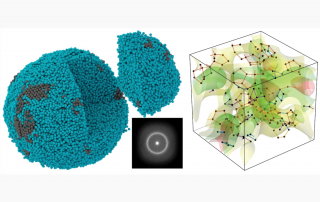
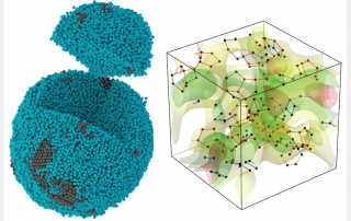
Electron microscopy is a high-resolution suite of characterization techniques used in the physical and biological sciences. By accelerating electrons to relativistic speeds (i.e. 0.5c) such that their characteristic wavelengths are 100,000 times smaller than visible light, one can perform high-resolution imaging down to the atomic scale. Furthermore, by implementing an array of diffraction and spectroscopic methods, electron microscopy can be used to decipher the nanoscale structure and composition of materials. This tutorial will begin by introducing electron microscopy and highlighting its advantages and disadvantages over visible and x-ray characterization techniques. Following this, the applications of electron microscopy will be summarized, with a specific focus on the cutting-edge experiments being performed by members of the STROBE community.