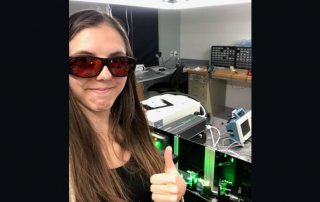
News

Home \ News

Congrats to Laura Waller for Being Selected as an American Institute for Medical and Biological Engineering (AIMBE) Fellow in 2019

Laura Waller has been selected as a 2019 AIMBE Fellow. The College of Fellows – 2,000 individuals who are outstanding bioengineers in academia, industry, clinical practice, and government. These leaders in the field have distinguished themselves through their contributions in research, industrial practice and/or education. Fellows are nominated each year by their peers and represent the top 2% of the medical and biological engineering community. They are considered the life-blood of AIMBE and work towards realizing AIMBE’s vision to provide medical and biological engineering innovation for the benefit of humanity.

Going Cold: The Future of Electron Microscopy

Researchers use electron microscopy to produce high-resolution images at the atomic scale of everything from composite nanomaterials to single proteins. The technology provides invaluable information on the texture, chemistry, and structure of these materials. Research over the past few decades has focused on achieving higher resolutions: being able to image materials at progressively finer levels with more sensitivity and contrast. But what does the future hold for electron microscopy?

Namrata Ramesh Receives Rhodes Scholarship

Berkeley Physics congratulates physics major Namrata Ramesh on being awarded a Rhodes Scholarship.

Namrata is in her senior year, pursuing a Physics (Honors) degree. Her senior thesis, supervised by Professor Naomi Ginsberg, involves understanding the dynamics of self-assembly of gold nanocrystal superlattices using optical and x-ray scattering techniques. She has also worked on studying the trajectories of electrons in manganese doped halide perovskites using Monte Carlo simulations. At Oxford, she hopes to continue investigating the origins of intriguing phenomena in promising photovoltaic materials by being at the interface of experimental and computational physics. Namrata is also very passionate about diversity in STEM fields and multimedia storytelling and has combined both interests by starting “The STEMinist Chronicles”, an organization that currently uses photo essays to tell the stories of women in STEM.

Congrats to Namrata Ramesh for being Awarded a Rhodes Scholarship

Namrata is a senior at the University of California, Berkeley, pursuing a Physics (Honors) degree. Her senior thesis, supervised by Professor Naomi Ginsberg, involves understanding the dynamics of self assembly of gold nanocrystal superlattices using optical and x-ray scattering techniques. She has also worked on studying the trajectories of electrons in manganese doped halide perovskites using Monte Carlo simulations.

Congrats to Rafael Piestun for Receiving the Lab Venture Challenge Award for Ultrathin Endoscopes

Rafael Piestun for received the 2019 Lab Venture Challenge Award for Ultrathin Endoscopes. Through the Lab Venture Challenge, Venture Partners at CU Boulder funds the top innovations showing high commercial potential, a clear path to a compelling market, and strong scientific support. In just the last 3 years, more than 20 commercially promising projects at CU Boulder have received funding through this program. Those same awards are associated with 16 new startup companies, with many having already raised further capital, demonstrating a powerful way to advance innovative research and translate it into impactful business ventures.

Congrats to Chris Regan and William Hubbard for Receiving the 2019 Microscopy Today Innovation Award

The editors of Microscopy Today congratulate the winners of the tenth Microscopy Today Innovation Award competition. The ten innovations advance microscopy in several areas: light microscopy, electron microscopy, and scanning probe microscopy. These innovations will make microscopy and microanalysis more powerful, more productive, and easier to accomplish.Secondary Electron Electron-BeamInduced-Current (SEEBIC) Imaging University of California at Los Angeles Developers: Chris Regan and William Hubbard. While intimately related to prior electron-beaminduced-current (EBIC) methods in the SEM, secondary electron electron-beaminduced-current (SEEBIC) imaging is qualitatively and quantitatively different. What makes the SEEBIC system new is that both the secondary electron (SE) and hole signals are detected in a scanning transmission electron microscope (STEM). SEEBIC differs from traditional EBIC in several ways. The measuring circuits are wired differently. In the former case the end of the device remote from the transimpedance amplifier is extremely high impedance, while in the latter it is tied to a low impedance (usually ground) to allow charge neutralization. While traditional EBIC imaging is sensitive to holes, it only generates contrast in regions where the sample supports an electric field that will separate electron-hole pairs. In most samples such regions are special and localized, for example, in a p-n junction. Thus, most of the sample generates no contrast when imaged with traditional EBIC. SEEBIC, on the other hand, is an inevitable consequence of imaging a thin specimen with an energetic electron beam, and SEEBIC imaging generates contrast everywhere in a sample. SEEBIC imaging has not been demonstrated previously for a couple reasons. First, the typical SEM sample is electron-opaque, and primary beam absorption produces a large background; thus, the SEEBIC signal is buried in the noise of the traditional SEM EBIC apparatus. This background is largely absent in the electron-transparent samples used in STEM. Secondly, the secondary electron (SE) yield drops with increasing beam energy; therefore, the SE signal is even smaller in a 200 kV STEM than in a 30 kV SEM. Detection of the signal requires a current measuring system that is low-noise and protected from electromagnetic interference (1 pA EBIC corresponds to ∼6,000 electrons in a 1 ms dwell time). SEEBIC is sensitive to electric potential, electric field, work function, conductivity, and temperature, and it can probe these quantities with atomic resolution in a modern STEM. STEM SEEBIC can image a functioning resistive random access memory (RRAM). For example, in a HfO2-based RRAM, the conducting filament is thought to consist of oxygen vacancies. Oxygen vacancies are basically invisible in a standard STEM image, but they give excellent contrast when viewed with STEM EBIC imaging.

KMLabs QM Quantum Microscope an R&D 100 Awards winner

Winners of the R&D 100 Awards have been announced by R&D World magazine and its new parent company, WTWH Media, LLC.

We are proud to announce that the QM Quantum Microscope™ is one of this year’s winners.  The QM Quantum Microscope builds on the company’s world leading technology in high harmonic generation to enable a range of techniques including coherent diffraction imaging, photoemission, pump-probe spectroscopy, and EUV metrology.

Primary Contributors to the project include:

JILA: Michael Tanksalvala, Yuka Esashi, Christina Porter, Michael Gerrity, Ting Liao, Margaret Murnane

KMLabs: Seth Cousin, Daisy Raymondson, Brennan Peterson, Henry Kapteyn

“Microscopes illuminated by coherent extreme UV beams are extremely sensitive to structure, composition and function at the nanoscale. They represent an entirely new class of lab scale microscope, with unique capabilities that are critical for future semiconductor, energy, solid state chemistry, and quantum devices.’ Henry Kapteyn, CTO.

About KMLabs: KMLabs is the only commercial provider for comprehensive, end-to-end research systems that leverage ultrafast pulses of extreme UV and soft X-ray light for a variety of experiments. The QM Quantum Microscope™ builds on the company’s world leading technology in high harmonic generation to enable a range of techniques including coherent diffraction imaging, photoemission, pump-probe spectroscopy, and EUV metrology. In addition, KMLabs continues to pioneer the development and engineering of standalone short wavelength sources including the Y-Fi VUV laboratory-based vacuum ultraviolet femtosecond laser source, and the Pantheon™ platform, a pulsed EUV source-beamline to generate and deliver EUV photons to user-supplied experimental stations.

2019 R&D 100 Award winners unveiled!

Winners of the R&D 100 Awards have been announced by R&D World magazine and its new parent company, WTWH Media, LLC. “This awards program is so well recognized across the R&D community. Being named as one of the R&D 100 is an incredible honor,” said Paul J. Heney, Vice President, Editorial Director for R&D World. “These 100 winning products and technologies are the disruptors that will change industries and make the world a better place in the coming years.”

Analytical/Test Category:

QM Quantum Microscope – Next Generation Microscopy & Analysis
KMLabs, Inc.
JILA at the University of Colorado, the STROBE center

Congrats to Michael Tanksalvala, Yuka Esashi, Christina Porter, Michael Gerrity, Ting Liao, Margaret Murnane (JILA), Seth Cousin, Daisy Raymondson, Brennan Peterson, and Henry Kapteyn (KMLabs) for Receiving the R&D 100 Award for the QM Quantum Microscope

Winners of the R&D 100 Awards have been announced by R&D World magazine and its new parent company, WTWH Media, LLC. “This awards program is so well recognized across the R&D community. Being named as one of the R&D 100 is an incredible honor,” said Paul J. Heney, Vice President, Editorial Director for R&D World. “These 100 winning products and technologies are the disruptors that will change industries and make the world a better place in the coming years.”

Analytical/Test Category:

QM Quantum Microscope – Next Generation Microscopy & Analysis
KMLabs, Inc.
JILA at the University of Colorado, the STROBE center

Go to Top